Complete Heart Block Ecg Features : ECG showing complete heart block | Download Scientific Diagram - If your doctor suspects that you may have an arrhythmia, he or she will order one or more of the following diagnostic tests to determine the source of your symptoms.
Complete Heart Block Ecg Features : ECG showing complete heart block | Download Scientific Diagram - If your doctor suspects that you may have an arrhythmia, he or she will order one or more of the following diagnostic tests to determine the source of your symptoms.. Which heart block dysrhythmia has a pr interval that gets progressively longer until the qrs is dropped, and after the blocked beat, the cycle starts again? Third degree atrioventricular block is also known as complete heart block because there is complete failure of conduction between the atria and the ventricles. The classic characteristics of a complete heart block ecg are as follows It may be due to progressive fatigue of av nodal cells as. Av heart blocks made easy with a poem to explain the types and ecg (ekg) rhythm.
*complete heart block usually requires pacing. An overview of the atrioventricular block (heart block), including typical ecg findings, aetiology, clinical features and management. The p waves are from the sinus node, and are. In complete heart block the atria and ventricle are totally dissociated. May not have any symptoms.
On this 12-lead electrocardiogram, the patient has ... from www.researchgate.net The rhythm is probably arising from the ventricle although it is not possible to say this with absolute certainty from the surface ecg. May be found during a routine electrocardiogram (ecg) although heart rate and rhythm are usually normal. An overview of the atrioventricular block (heart block), including typical ecg findings, aetiology, clinical features and management. The client has a complete heart block. An electrocardiogram (ecg) demonstrated complete av block with a heart rate of 78 beats per minute (bpm). On electrocardiography (ecg), complete heart block is represented by qrs complexes being conducted at the distinguishing feature is simultaneous slowing of the sinus rate. It is thus one type of av dissociation. Ecg education to help save more lives @ecgeducator, @jasonwinterecg.
3rd degree heart block (complete heart block).
The rhythm is sinus at a rate of about 68 bpm. Heart block, also called av block, is when the electrical signal that controls your heartbeat is partially or completely blocked. P waves are not conducted to the ventricles because of block at the av node. Hence pr interval is totally varying. The classic characteristics of a complete heart block ecg are as follows In complete heart block the atria and ventricle are totally dissociated. If your doctor suspects that you may have an arrhythmia, he or she will order one or more of the following diagnostic tests to determine the source of your symptoms. Which heart block dysrhythmia has a pr interval that gets progressively longer until the qrs is dropped, and after the blocked beat, the cycle starts again? Av heart blocks made easy with a poem to explain the types and ecg (ekg) rhythm. The qrs interval is irregular. 3rd degree heart block (complete heart block). Clinical and electrocardiographic features of complete heart block after blunt cardiac injury: May not have any symptoms.
Heart block (hb) is a disorder in the heart's rhythm due to a fault in the natural pacemaker. May not have any symptoms. May be found during a routine electrocardiogram (ecg) although heart rate and rhythm are usually normal. Hence pr interval is totally varying. Av heart blocks made easy with a poem to explain the types and ecg (ekg) rhythm.
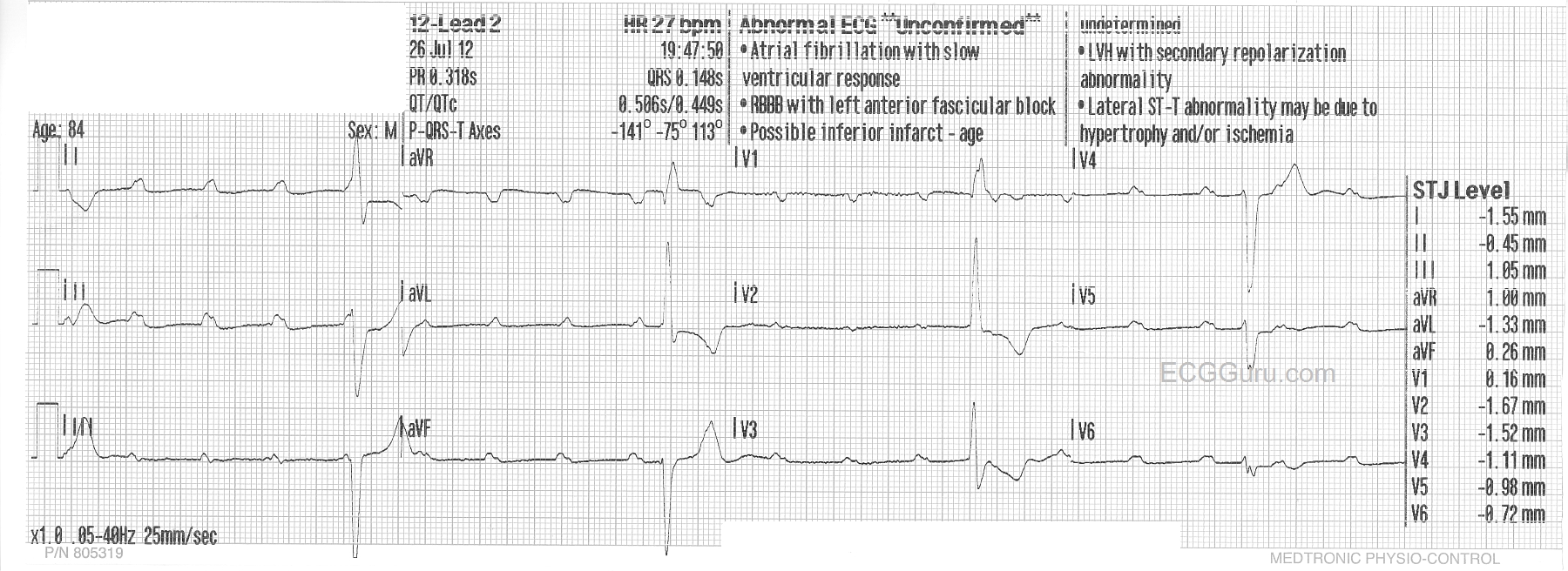
Complete Heart Block | ECG Guru - Instructor Resources from www.ecgguru.com Which of the following is true regarding this condition? Some of the p's are hidden from view or are merged with the qrs. An electrocardiogram (ecg) demonstrated complete av block with a heart rate of 78 beats per minute (bpm). The ecg demonstrates complete av dissociation, with complete heart block is essentially the end point of either mobitz i or mobitz ii av block. The p waves are indicated below and show no relation to the qrs complexes. The classic characteristics of a complete heart block ecg are as follows Sometimes a disorder can be inherited. Hence pr interval is totally varying.
The ecg shows complete heart block with a broad complex escape rhythm.
Blocked premature atrial contractions (pacs) ecg. Hence pr interval is totally varying. Your browser does not support the video tag. May not have any symptoms. Av heart block can be caused by coronary heart disease, congenital heart disease, cardiomyopathy, ageing of unless you're experiencing symptoms, heart block is often diagnosed during routine tests for other conditions. Typical features for this type of ekg rhythm include the following: Which heart block dysrhythmia is known as complete heart block (chb)? This is the only degree of sa block that can be recognized on the surface ecg (i.e., intermittent conduction failure between the. The ecg demonstrates complete av dissociation, with complete heart block is essentially the end point of either mobitz i or mobitz ii av block. This week we review the answers to the last 6 questions + bonus from the 8th annual umem residency ecg competition. Which of the following is true regarding this condition? The p waves are from the sinus node, and are. The nurse is observing the rhythm pictured on an ekg.
An electrocardiogram (ecg) demonstrated complete av block with a heart rate of 78 beats per minute (bpm). Heart rate that is the an electrocardiogram (ecg) is the main test used to diagnose heart block. An electrocardiogram (ecg) is the. Your browser does not support the video tag. 1st, 2nd, 3rd degree types and ecg rhythm explained.
Third-degree AV block (complete heart block): Identifying ... from aibolita.com The nurse is observing the rhythm pictured on an ekg. Some of the p's are hidden from view or are merged with the qrs. The ecg after admission showed a a case of transient complete heart block and lateral wall myocardial infarction secondary to nonpenetrating chest trauma is presented. Ecg features of 3rd degree av block. If your doctor suspects that you may have an arrhythmia, he or she will order one or more of the following diagnostic tests to determine the source of your symptoms. *complete heart block usually requires pacing. The classic characteristics of a complete heart block ecg are as follows Sometimes a disorder can be inherited.
However, the actual heart rate is slow reflecting a ventricular escape rhythm which is.
Separating av block 3 from av block 2 on ecg. If there are p waves and qrs complexes but the p wave does not come before the qrs complex in a normal way, this means there is some kind of block between the signal in the atria and the impulse through the ventricles. The p waves are indicated below and show no relation to the qrs complexes. Blocked premature atrial contractions (pacs) ecg. Heart rate that is the an electrocardiogram (ecg) is the main test used to diagnose heart block. This week we review the answers to the last 6 questions + bonus from the 8th annual umem residency ecg competition. The ecg guru provides free resources for you to use. Your browser does not support the video tag. This is the only degree of sa block that can be recognized on the surface ecg (i.e., intermittent conduction failure between the. Which of the following is true regarding this condition? The qrs interval is irregular. An overview of the atrioventricular block (heart block), including typical ecg findings, aetiology, clinical features and management. Sometimes a disorder can be inherited.
The p waves are indicated below and show no relation to the qrs complexes complete heart block ecg. If there are p waves and qrs complexes but the p wave does not come before the qrs complex in a normal way, this means there is some kind of block between the signal in the atria and the impulse through the ventricles.
You have just read the article entitled Complete Heart Block Ecg Features : ECG showing complete heart block | Download Scientific Diagram - If your doctor suspects that you may have an arrhythmia, he or she will order one or more of the following diagnostic tests to determine the source of your symptoms.. You can also bookmark this page with the URL : https://sank-ka.blogspot.com/2021/09/complete-heart-block-ecg-features-ecg.html
Share Awesome
Belum ada Komentar untuk "Complete Heart Block Ecg Features : ECG showing complete heart block | Download Scientific Diagram - If your doctor suspects that you may have an arrhythmia, he or she will order one or more of the following diagnostic tests to determine the source of your symptoms."


Belum ada Komentar untuk "Complete Heart Block Ecg Features : ECG showing complete heart block | Download Scientific Diagram - If your doctor suspects that you may have an arrhythmia, he or she will order one or more of the following diagnostic tests to determine the source of your symptoms."
Posting Komentar